Transforming
Cancer Diagnostics with AI
Chaim Linhart, Ph.D. Co-founder & CTO, Cobi Reouven, Principal Software Engineer
Through a strategic partnership with Maccabi Healthcare Services, an Israeli HMO that works with the Philips IntelliSite Pathology Solution, we have access to millions of pathology slides, reports, and medical records. This data is essential for developing our medical-grade algorithms, which require high-quality images and scans. Ibex uses the Philips Pathology Software Development Kit (SDK) in three application areas. One is patch extraction, where we read small image regions in various magnifications and from multiple locations. These patches are used for training our classification models (CNNs) and for inferencing, i.e., diagnosing new slides using the trained models. Another use-case is image conversion. We use the Pathology SDK to convert the iSyntax format files to TIFF files so that slides can be viewed in third party tools (e.g., slide viewers) that do not yet have native iSyntax read capabilities, and for reduced storage footprint available for TIFF files, in situations where compression is acceptable. The third use case is extracting metadata of a slide, for example, its dimensions, the magnification levels in which the slide was scanned, or its envelopes, which are the areas that the scanner scanned within the slide. For whole slide images, we work with either the native iSyntax format, exported TIFF (a manual option available on the Philips IntelliSite Pathology Solution), or images that are converted to TIFF using sample code, which is part of the Pathology SDK. It should be mentioned that exported TIFF files go through image processing for conversion and further compression, which can take a fair bit of time. More importantly, iSyntax files, while having a larger file size, have medical-grade image quality. We embedded the Pathology SDK in our code - we wrote a patch extraction layer that hides the extraction implementation from the applicative code. This layer can use either the SDK for reading iSyntax files or the OpenSlide library for reading TIFF files. In our tests, we saw very similar accuracy results with both models, which means that we did not compromise accuracy by using compressed files. However, in terms of runtime, we noticed that the SDK reads large patches from iSyntax files much faster than OpenSlide reads TIFF files, with a more noticeable impact in larger patches. In our application pipeline, specifically the inference, we read vast regions, so for us, the performance impact with native iSyntax image format is significant.
Ibex is a startup based in Israel, developing Artificial Intelligence (AI) based software tools that help pathologists diagnose cancer more accurately, more efficiently, and more objectively. We were the first to develop and deploy an AI-based solution used in routine clinical diagnosis of prostate core needle biopsies - The Ibex Second Read System. The system alerts on potential diagnostic errors, thus providing a safety net for pathologists and patients. Ibex brings the AI revolution to pathology laboratories and improves cancer care.
Share on social media
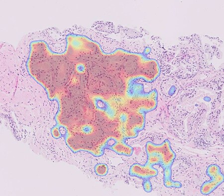
This image was taken from a cancer case diagnosed initially as benign by a pathologist, but following an alert by the Ibex Second Read System was re-diagnosed as cancerous resulting incorrect treatment for the patient. The red area includes cancer that was missed in the first diagnosis and was then re-diagnosed.