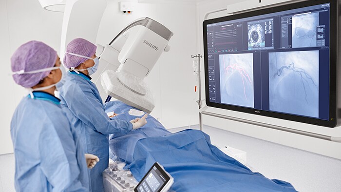
Cerebral aneurysm treatment
See clearly and navigate efficiently when treating cerebral
aneurysms
aneurysms
Demonstrated results
100%
of users
found that controlling SmartCT is intuitive and easy to learn[1] and increases clinical confidence with advanced 3D imaging, visualization and measurement tools.
88%
of users
believe they can have more focus on their patient - thanks to full tableside control with the touchscreen module[1]
75%
dose reduction in neuro DSA[2]
with ClarityIQ compared to a system without ClarityIQ. Its automatic motion compensation removes skull and motion artifacts, which is key when placing small devices at the base of the skull
Image gallery
- Toggle view
Featured solutions for cerebral aneurysm treatment

Azurion 7 B20/15 Image Guided Therapy System Biplane
High-quality neuro-specific lateral detector can be positioned close to the patient’s head while providing full brain coverage. Secure, fast lateral gantry parking allows seamless switch between 2D and 3D image acquisition. Frontal arc 135° position enables optimal head-end access to the patient.

SmartCT Vaso
SmartCT Vaso enables visualization of both low contrast soft tissue, contrast enhanced vessels and high contrast objects such as flow diverters, stents, intrasaccular devices and coils in one view which allows users to assess device deployment.

SmartCT Soft Tissue
Helical*
Improved neuro CT-like cone beam CT images (CBCT) to identify ischemic changes in the angio suite. An advanced protocol with dual-axis acquisition trajectory and improved reconstruction software results in improved image appearance, compared to conventional CBCT acquisition techniques.

SmartCT Dual Viewer*
Simultaneously visualize two 3D datasets of different sizes--acquired at different times during the procedure--and overlay them to create fusion images to support assessment and diagnosis without breaking sterility by using the touchscreen module or mouse and keyboard.

SmartCT Roadmap
Enhance visualization of overlapping vessels to support precise navigation of guidewire and catheter through complex vasculature. Offers high-level precision with real-time compensation for gantry, table and small patient movements. Customize Roadmap Pro to show advancement during coil placement.

SmartCT Angio
Improve visibility of tortuous or complex anatomy by generating a complete high-resolution 3D visualization of cerebral vasculature from a single rotational angiography run – all controlled via the touchscreen at tableside.

AneurysmFlow
Assessing the impact on blood flow by an embolization device--such as a flow diverter--right after deployment is crucial. AneurysmFlow cerebral aneurysm flow quantification is designed to provide relevant information based on quantification of blood flow changes.
"The VasoCT multiplanar understanding is extremely easy to use and to use it directly in the angio suite."
Prof. Vincent Costalat
Departement head of Therapeutic & Diagnostic Neuroradioogy, Centre Hospitalier Universitaire de Montpellier, France."The system is like a very mindful assistant in the background, and I do not have to concentrate on other things than the patient.”
PD Dr. Tobias Boeckh - Behrens
Deputy managing director of the Department of diagnostic and interventional Neuroradiology , Head of Neurointervention , University Hospital Rechts der Isar, Munich, Germany
Technologies and innovations
Disclaimer:
*SmartCT R3.0 is subject to regulatory clearance and may not be available in all markets. Contact your sales representative for more details.
Product availability is subject to country regulatory clearance. Please contact your local sales representative to check the availability in your country.
[1] Market Research Group reports ‘16-’17.
[2] Söderman M, Holmin S, Andersson T, Palmgren C, Babic D, Hoornaert B. Image noise reduction algorithm for digital subtraction angiography: clinical results. Radiology. 2013 Nov;269(2):553-60. The results of the application of dose reduction techniques will vary depending on the clinical task, patient size, anatomical location and clinical practice. The interventional radiologist assisted by a physicist as necessary has to determine the appropriate settings for each specific clinical task.


