Peripheral IVUS
Advanced visualization for deeper insights with Philips IVUS
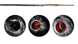
IVUS is a catheter-based imaging technology that allows visualization of vessels from the inside out. Cross-sectional images help assess presence and extent of disease, plaque geometry and morphology, guide wire position during lesion crossing, and stent position post-treatment. Only market-leading Philips IVUS portfolio offers a plug-and-play platform for Peripheral IVUS and a full line of catheters for arterial and venous interventions.
Optimize outcomes with Philips IVUS
79%
Randomized controlled trial demonstrates that IVUS changed treatment plans in 79% of arterial cases and 57% in venous cases, outlining more informed treatment plans for patients.
changes in treatment plans with IVUS1,2
27%
Reductions in major adverse arterial limb events3
Data from the largest ever real-world clinical analysis of over 700k peripheral arterial and peripheral venous patients on lower extremity peripheral intervention for IVUS, demonstrate a 27% reduction in risk for major adverse limb events including acute limb ischemia or major amputation.
28%
Reductions in repeat venous interventions, hospitalization or death4
Data from the largest ever real-world clinical analysis of over 700k peripheral arterial and peripheral venous patients on lower extremity peripheral intervention for IVUS, demonstrate a 28% risk reduction for repeat intervention, hospitalization, or death.
1st
ever
Global consensus guidance on IVUS5
40 cross-specialty experts achieved consensus for the appropriate use of IVUS in peripheral vascular disease interventions. The new recommendations aim to improve quality care. IVUS is recommended for lower extremity arterial and venous revascularization procedures to guide clinical decisions.
Features

Plays an essential role in disease assessment
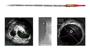
IVUS imaging helps aid disease diagnosis, including plaque burden percentage, lesion location and morphology, calcium volume, and the presence of thrombus. It also provides analysis of crucial parameters—like luminal cross-sectional measurements.

Supports treatment decisions during intervention
IVUS provides real-time diagnostic imaging for peripheral artery disease and venous disease. It may also guide clinicians to the correct angioplasty technique for a patient’s individual needs, help assess intervention effectiveness, and assist in endovascular device delivery. The imaging modality also helps clinicians determine the size of the device needed for the best outcome.

Confirms treatment results and optimizes outcomes
IVUS imaging helps to confirm treatment results, including diagnosing dissections, the completeness of treatment, and the apposition and expansion of stent placement.

Minimizes procedural complications
IVUS-Guided Re-Entry Catheter – Pioneer Plus has demonstrated a 95% to 100% procedural success rate in subintimal angioplasty procedures.6 The guidance and direction provided by on-board IVUS imaging has the potential to minimize procedural complications.7

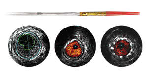
Assess stent apposition with ChromaFlo
On select catheters*, ChromaFlo highlights blood flow red to ease assessment of stent apposition, lumen size, and more. Appropriate for peripheral and coronary vessels, including left main, bifurcations, superficial femoral artery and iliac, it is designed to make lumen size and stent apposition instantly recognizable and helps identify branches, dissections, and plaque in bifurcations.

Reduces radiation to patient and interventionalist
Philips IVUS may reduce the use of angiograms, thereby reducing exposure to procedural radiation. In addition to the patient, interventionalists are also exposed to significant radiation, compounded by the number of procedures they perform.
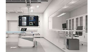
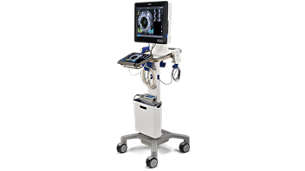
Peripheral IVUS technology is available on
“A moment that really changed a patient's outcome using IVUS for me was a patient that was told had adequate perfusion to their foot based on outside imaging and workups."
Kumar Madassery, MD
Interventional Radiologist
Rush University Medical Group
Chicago, IL“I had a patient where I had a treatment plan set aside after seeing the initial angiograms and then once I did the IVUS recognized that there was more to it, seeing thrombus inside the vessel, which I did not appreciate with two-dimensional imaging.”
Jon George, MD, MBA
Interventional Cardiologist
Penn Medicine
Philadelphia, PA Philadelphia, PA
“I cant think of one patient, I can think of hundreds of patients. The beauty of IVUS is that we can individualize our treatment algorithms for that patient.”
Pradeep Nair, MD, MBA
Interventional Cardiologist
Cardiovascular Institute of the South
Houma, LA

IVUS reimbursement now available!
IVUS reimbursement is now available for medically necessary patients in all sites of service. Contact the Philips Health Economics team at IGTDReimbursement@philips.com to learn more.
Brochures
- Brochures
-
Footnotes
*ChromaFlo is available on select peripheral IVUS catheters including: OTW Digital IVUS Catheter – Reconnaissance PV .018, RX Digital IVUS Catheter – Visions PV. 018 RX, Visions PV .014P RX, and IVUS-Guided Re-Entry Catheter – Pioneer Plus [2] Allan R, Puckridge P, Spark J, et al. The Impact of Intravascular Ultrasound on Femoropopliteal Artery Endovascular Interventions. J Am Coll Cardiol Intv. 2022 Mar, 15 (5) 536–546. https://doi.org/10.1016/j.jcin.2022.01.001 [3] Divakaran S, Parikh SA, Hawkins BM, et al. Temporal Trends, Practice Variation, and Associated Outcomes With IVUS Use During Peripheral Arterial Intervention. JACC Cardiovasc Interv. 2022;15(20):2080-2090. doi:10.1016/j.jcin.2022.07.050 [4] Divakaran S, Meissner MH, Kohi MP, et al. Utilization of and Outcomes Associated with Intravascular Ultrasound during Deep Venous Stent Placement among Medicare Beneficiaries. J Vasc Interv Radiol. 2022;33(12):1476-1484.e2. doi:10.1016/j.jvir.2022.08.018 [5] Secemsky EA, Mosarla RC, Rosenfield K, et al. Appropriate Use of Intravascular Ultrasound During Arterial and Venous Lower Extremity Interventions. JACC Cardiovasc Interv. 2022;15(15):1558-1568. doi:10.1016/j.jcin.2022.04.034 [6] Al-Ameri, H et al. Peripheral Chronic Total Occlusions Treated with Subintimal Angioplasty and a True Lumen Re-Entry Device. Journal of Invasive Cardiology. 2009; 21(9): 468-472. [7] Saketkhoo RR, Razavi MK, Padidar A, Kee ST, Sze DY, Dake MD. Percutaneous bypass: subintimal recanalization of peripheral occlusive disease with IVUS guided lumen re-entry. Tech Vasc Interv Radiol. 2004; 7: 23-27.
[1] Gagne PJ, Tahara RW, Fastabend CP, et al. Venography versus intravascular ultrasound for diagnosing and treating iliofemoral vein obstruction. J Vasc Surg Venous Lymphat Disord. 2017;5(5):678-687. doi:10.1016/j.jvsv.2017.04.007
Always read the label and follow the directions for use. Philips medical devices should only be used by physicians and teams trained in interventional techniques, including training in the use of this device.Products subject to country availability. Please contact your local sales representative.
©2024 Koniklijke Philips N.V. All rights reserved. Trademarks are the property of Koninklijke Philips N.V. or their respective owners. Philips reserves the right to change product specifications without prior notification.

