Complicated assessments made easy
Due to an aging population as well as increases in obesity and sports activity, joint surgeries are on the rise. Perform a wide variety of tasks, such as assessment and reconstruction, thanks to the orthopedics application suite on IntelliSpace Portal. The tools are designed for even the most challenging musculoskeletal cases.
A comprehensive approach to orthopedics
Take a closer look at cases involving bone and cartilage. CT Acute MultiFunctional Review, for example, includes an MSK and surgical planning stage. CT Bone Mineral Analysis, previously only available on Philips Extended Brilliance Workspace, helps you track and manage degenerative and metabolic bone diseases.
- Acute Multi-Functional Reivew (AMFR)
-
CT Acute MultiFunctional Review (AMFR)
One application for the assessment of selected anatomies
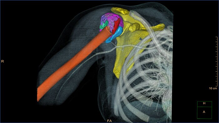
CT Acute MultiFunctional Review (AMFR) provides dedicated tools for findings detection, visualization and assessment of vessels, bones and spine anatomies in 2D and 3D CT images.
Benefits
- Automatic navigation-path for calculation of the spinal cord as well as automatic detection and labeling of spine vertebrae and discs.
- Bones segmentation using an interactive segmentation tool to create a workspace for virtual repositioning of individual bone segments.
- Provides segmentation, editing and measurement tools for vascular analysis.
- Predefined layouts per anatomical area: head, chest, abdomen, spine and extremities.
- Bone Mineral Anlysis (BMA)
-
CT Bone Mineral Analysis (BMA)
Track degenerative and metabolic bone disease
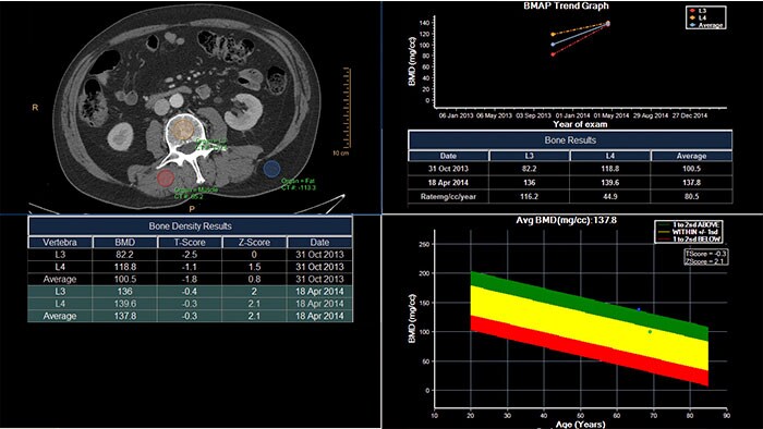
CT Bone Mineral Analysis (BMA) is designed to measure bone density in one or multiple time points. Using an internal reference method*, the application reduces reproducibility errors in multiple time point measurements and provides T- and Z- scores which help physicians assess the risk of osteoporosis.
Benefits
- The user can compare a patient’s results to several reference populations.
*Muller DK, et al., Phantom-less QCT BMD system as screening tool for osteoporosis without additional radiation. Eur J Radiol. 2011; 79(3):375–81.
- Spectral Light Magic Glass
-
CT Spectral Light Magic Glass
Review spectral data in a range of not spectral-enhanced CT applications
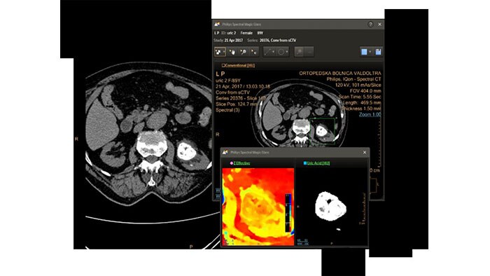
Allows retrospective use of spectral data that was saved in a series of spectral base images (SBI).
The fast launch of LMG allows review and identification of the most relevant results to be launched into the application for further analysis.
Benefits
- The option is available from the following applications: Brain Perfusion, Functional CT, Liver Analysis, PAA, TAVI, Acute Multifunctional Review, Virtual Colonoscopy.
- Spectral Magic Glass can be launched only for CT images or images created on the Philips IQon Spectral CT.
- Spectral Magic Glass on PACS
-
CT Spectral Magic Glass on PACS*
IQon Spectral CT Functionality
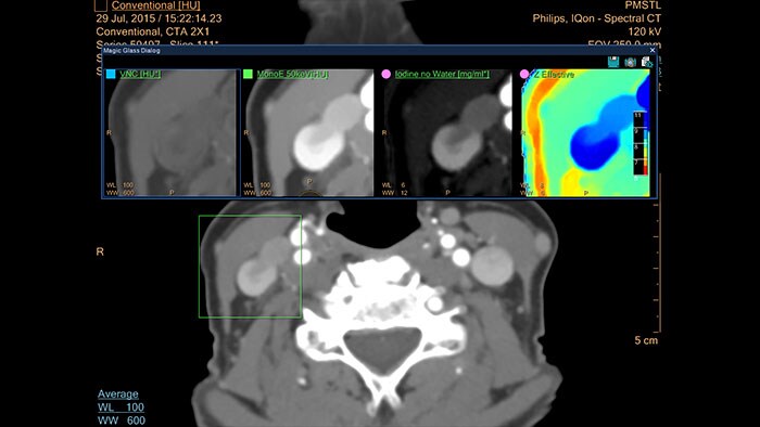
IQon Spectral CT is the only scanner to offer CT Spectral Light Magic Glass and CT Spectral Magic Glass on PACS, helping radiologists review and analyze multiple layers of spectral data at once, including on their PACS.
Benefits
- On-demand simultaneous analysis of multiple spectral results for an Region Of Interest (ROI).
- Integrates into a health system’s current PACS setup for certain PACS vendors.
- Spectral results viewable, during a routine reading.
- Enterprise-wide spectral viewing and analysis allows access to capabilities virtually anywhere in the organization.
* Standard with the CT Spectral option on IntelliSpace Portal.
- Spectral Viewer
-
CT Spectral Viewer
IQon Spectral CT* Functionality
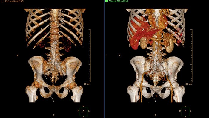
The spectral viewer is optimized for analysis of spectral data sets from the IQon Spectral CT Scanner. Obtain a comprehensive overview of each patient quickly and easily, quantify quickly, and assist in diagnosis. It is designed to accommodate general spectral viewing needs with additional tools to assist in CT images analysis.
Benefits
- Enhances the conventional image by overlaying an iodine map.
- Visualization of virtual non-contrast images.
- Images at different energy levels (40-200 keV).
- Switching to various spectral results can be done through a viewport control.
- Manage presets to create user/site-specific presets.
- Lesion characterization using scatter plots.
- Tissue characterization using attenuation curves.
* IQon CT reconstruction provides a single DICOM entity containing sufficient information for retrospective analysis - Spectral Base Image (SBI). SBI contains all the spectrum of spectral results with no need for additional reconstruction or post-processing. Spectral applications are creating different spectral results from SBI.
- 3D Modeling
-
3D Modeling
Streamlined modeling workflow
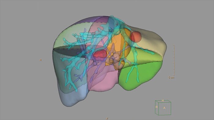
Allows to view volumetric images of anatomical structures, perform segmentation, edit and combine segmented elements (tissues) into a 3D model.
Benefits
- Studies of CT & MR can be used for creating a single 3D model of the same patient. The application provides tools that allow the user to align between the volumes of interest in the images.
- 3D Modeling batches files can be easily exported in standard formats such as STL, with the option to also provide a 3D PDF as an additional means for results sharing with 3D printing or other services* .
- The user may determine the information related to the exported elements of the 3D model such as smoothness and output mesh size.
- Contours can also be exported as RT Structures.
*3D models are not intended for diagnostic use.
- Cartilage Assessment
-
MR Cartilage Assessment
Visualize cartilage structures
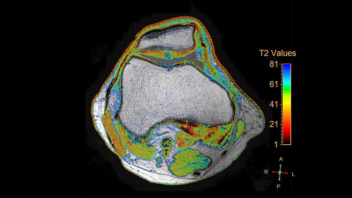
Enables the visualization of cartilage structures integrated with color-coded T2 maps. Positioning of cartilage-shaped, layered region of interest is used to assess variation of T2 values across the cartilage depth to determine the degradation of the cartilage.
Benefits
- The MR Cartilage Assessment application features a task-guided workflow for the quantitative analysis of T2 relaxation time to support cartilage assessment and disease status.
- The application provides segmentation tools, allowing measurements of cartilage layers and segments.
- T2 values are numerically and graphically displayed per layer and segment.
- Echo Accumulation
-
MR Echo Accumulation
Optimizing image contrasts for multi-echo MR data
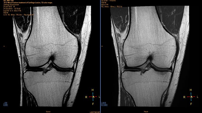
MR Echo Accumulation is used to perform pixelwise echo accumulations for imaging series with multiple echoes. MR Echo Accumulation enables the calculation of new images based on the selected sum of echo times of series with multiple echoes. The processing provides interactive update of the results.
Benefits
- MR Echo Accumulation enables the calculation of new images based on the selected sum of echo times.
- Provides the ability to define the accumulation range using an interactive slider.
- Enables preview, save and analysis on the calculated new series.
